Etiketter

Summa sidvisningar

Leta i den här bloggen

fredag 8 november 2019

Insuliinireseptorin isoformien A ja B suhteen merkityksestä ihmisessä? (2018)

https://cardiab.biomedcentral.com/articles/10.1186/s12933-018-0675-2
Potential role of insulin receptor isoforms and IGF receptors in plaque instability of human and experimental atherosclerosis
Article metrics
Clinical complications associated with atherosclerotic plaques arise from luminal obstruction due to plaque growth or destabilization leading to rupture. We previously demonstrated that overexpression of insulin receptor isoform A (IRA) and insulin-like growth factor-I receptor (IGF-IR) confers a proliferative and migratory advantage to vascular smooth muscle cells (VSMCs) promoting plaque growth in early stages of atherosclerosis. However, the role of insulin receptor (IR) isoforms, IGF-IR or insulin-like growth factor-II receptor (IGF-IIR) in VSMCs apoptosis during advanced atherosclerosis remains unclear.
 --

Results

We observed a significant decrease of IRA/IRB ratio in human complicated plaques as compared to non-complicated regions. Moreover, complicated plaques showed a reduced IGF-IR expression, an increased IGF-IIR expression, and lower levels of α-SMA indicating a loss of VSMCs. In experimental atherosclerosis, we found a significant decrease of IRA with an increased IRB expression in aorta from 24-week-old BATIRKO; ApoE−/− mice. Furthermore, atherosclerotic plaques from BATIRKO; ApoE−/− mice had less VSMCs content and higher number of apoptotic cells. In vitro experiments showed that IGF-IR inhibition by picropodophyllin induced apoptosis in VSMCs. Apoptosis induced by thapsigargin was lower in IR−/− VSMCs expressing higher IGF-IR levels as compared to IRLoxP+/+ VSMCs. Finally, IRB VSMCs are more prone to thapsigargin-induced apoptosis than IRA or IRLoxP+/+ VSMCs.

Conclusions

In advanced human atherosclerosis, a reduction of IRA/IRB ratio, decreased IGF-IR expression, or increased IGF-IIR may contribute to VSMCs apoptosis, promoting plaque instability and increasing the risk of plaque rupture and its clinical consequences.

Background
Atherosclerosis is a chronic disease affecting large arteries that involves the formation of plaques containing vascular and inflammatory cells, lipids and extracellular matrix [1]. Its clinical complications arise from luminal obstruction due to plaque growth leading to vessel stenosis, and/or formation of unstable plaques that acutely rupture leading to an occlusive thrombus formation [2]. Vascular smooth muscle cells (VSMCs) play a main role in this process as they contribute to plaque growth in early stages, but favor plaque stability in advanced stages of atherogenesis [3].

  • SUOMENNOSTA:
  •   Insuliinin (Ins)  ja insuliininkaltaisten kasvutekijöiden  (IGFs)  signalointi välittyy hormoni-interaktioilla ja reseptoreina toimivat  insuliinireseptori (IR) ja  IGF-I reseptori (IGF-IR), jotka ovat  Tyrosiinikinaasireseptorien superperheen  II-alaluokan jäseniä.  Imettäväisissä alternatiivisella pleissauksellaa insuliinireseptorigeenistä tulee kaksi isoformia IR A ja IR B. IRB omaa  12 aminohapon lisäsekvenssin, jota koodaa exoni 11. Vaikka molemmat isoformit omaavat saman affiniteetin insuliinia kohtaan, niin IRA  ilmentää suurempaa affiniteettia insuliininkaltaisia kasvutekijöitä ( IGFs) kohtaan, erityisesti IGF-II-muotoa kohtaan.  Täten voidaan todeta,että IRB ensisijassa on assosioitunut metabolisiin ja  differentioiviin signalointeihin, kun taas IRA pääasiassa suosii solukasvua, proliferaatiota ja elossapysymistä. Lisäksi IGF-II-muoto voi sitoutua  omaan IGF-II-reseptoriin (IGF-IIR) suurella affiniteetilla. 
  • IGF-IIR on tyypiltään transmembraaninen glykoproteiini, jolla on  suuri affiniteetti  myös mannoosi-6-fosfaattiin, joten se voi sitoutua myös lysosomaalisiin entsyymeihin ja muihin kasvutekijöihin ja sytokiineihin.  Sillä onkin  hyvin dolumentoitu tehtävä solunsisäisessä lysosomaalisten entsyymien kuljetuksessa ja IGF-II:n puhdistamisessa verenkierrosta. Mutta IGF-IIR ei omaa tyrosiinikinaasiaktiivisuutta eikä se autofosforyloidukaan, vaan linkkiytyy G-proteiinivälitteisesti signaalin johtumisverkostoon ja täten  voi osallistua solukäytöksen säätelyyn.

The insulin and insulin-like growth factors (IGFs) signaling is mediated by hormone interaction with the insulin receptor (IR) and the IGF-I receptor (IGF-IR), which are members of subclass II tyrosine kinase receptor super-family [4, 5]. In mammals, alternative splicing of the IR gene gives rise to two isoforms: IRA and IRB [6]. Indeed, IRB has an additional 12-amino acid sequence encoded by the exon 11. Although both isoforms have similar affinity for insulin, IRA exhibits a higher affinity for IGFs, especially for IGF-II [7]. Thus, IRB is preferentially associated with metabolic and differentiating signals, whereas IRA mainly favors cell growth, proliferation and survival [8]. In addition to IR and IGF-IR, IGF-II binds IGF-II receptor (IGF-IIR) with high affinity. IGF-IIR is a type I transmembrane glycoprotein that also have high affinity for mannose-6-phosphate, and can therefore bind lysosomal enzymes and other growth factors and cytokines [9]. It plays a well-documented role in the intracellular transport of lysosomal enzymes and in clearance of IGF-II from the circulation. However, although IGF-IIR contains neither tyrosine kinase activity nor an autophosphorylation site, it does link to G-proteins providing a mechanism for signal transduction that may be involved in cell behavior regulation [10, 11].
In early atherosclerotic lesions, IGFs contribute to plaque growth by promoting VSMCs proliferation and migration [12]. In this regard, we previously demonstrated that overexpression of IGF-IR or IRA isoform during early atherosclerosis confers a proliferative and migratory advantage to VSMCs favoring atherosclerotic progression [13, 14]. In advanced stages, the imbalance between cell death and survival may substantially affect the cellularity and integrity of atherosclerotic lesions contributing to plaque instability. Unstable plaques that are prone to rupture have a thin fibrous cap with a decreased number of VSMCs and a dense infiltration of inflammatory cells [15, 16], as well as an increased apoptosis of VSMCs and macrophages [17]. IGF-I, through IGF-IR, has been reported to prevent atherosclerotic plaque instability by its mitogenic and antiapoptotic effects on VSMCs [18,19,20]. However, the role of IR isoforms or IGF-IIR in VSMCs apoptosis and thereby in plaque instability remains unknown. In the present study, we analyzed the expression of IR isoforms, IGF-IR and IGF-IIR, as well as VSMCs content in human carotid atherosclerotic plaques and in experimental models of atherosclerosis. Finally, we assessed the contribution of IR isoforms and IGF-IR to the apoptosis of murine aortic VSMCs lines.
  •  SUOMENNOSTA TÄMÄN TUTKIMUKSEN TULOKSISTA.Komplisoiduissa ateroskleroottisissa plakeissa on havaittu, että  IRA/IRB-suhde  on laskenut  ia IGF-IR on laskenut, IGF-IIR on  lisääntynyt ja  VSMC- vaskulaarisia  sileitä lihassoluja on  kadonnut.
Results

Decrease of IRA/IRB ratio and IGF-IR, increase of IGF-IIR, and loss of VSMCs in complicated human atherosclerotic plaques

torsdag 7 november 2019

Millä tyylillä insuliini signaloi insuliinireseptorille? (2013)

https://www.nature.com/articles/nature11781

How insulin engages its primary binding site on the insulin receptor

Nature volume 493, pages 241245 (10 January 2013) | Download Citation

Abstract
Insulin receptor signalling has a central role in mammalian biology, regulating cellular metabolism, growth, division, differentiation and survival1,2. Insulin resistance contributes to the pathogenesis of type 2 diabetes mellitus and the onset of Alzheimer’s disease3; aberrant signalling occurs in diverse cancers, exacerbated by cross-talk with the homologous type 1 insulin-like growth factor receptor (IGF1R)4. Despite more than three decades of investigation, the three-dimensional structure of the insulin–insulin receptor complex has proved elusive, confounded by the complexity of producing the receptor protein. Here we present the first view, to our knowledge, of the interaction of insulin with its primary binding site on the insulin receptor, on the basis of four crystal structures of insulin bound to truncated insulin receptor constructs.  

The direct interaction of insulin with the first leucine-rich-repeat domain (L1) of insulin receptor is seen to be sparse, the hormone instead engaging the insulin receptor carboxy-terminal α-chain (αCT) segment, which is itself remodelled on the face of L1 upon insulin binding.

 Contact between insulin and L1 is restricted to insulin B-chain residues. The αCT segment displaces the B-chain C-terminal β-strand away from the hormone core, revealing the mechanism of a long-proposed conformational switch in insulin upon receptor engagement. This mode of hormone–receptor recognition is novel within the broader family of receptor tyrosine kinases5.

 We support these findings by photo-crosslinking data that place the suggested interactions into the context of the holoreceptor and by isothermal titration calorimetry data that dissect the hormone–insulin receptor interface.

Together, our findings provide an explanation for a wealth of biochemical data from the insulin receptor and IGF1R systems relevant to the design of therapeutic insulin analogues.

Insuliinireseptorista ja insuliinista artikkeli heinäkuulta 2019

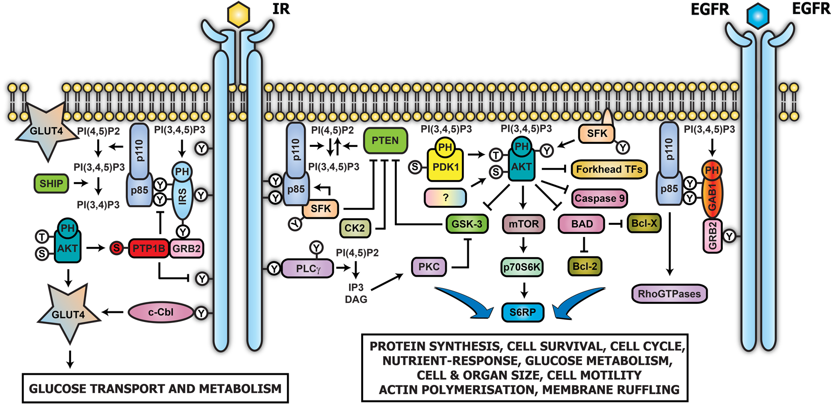
 Insuliinireseptorista eräs kuva: http://www.cellsignet.com/media/proteins/InsRPI3K-EGFRGab1.png


Insuliinista eräskuva: https://www.researchgate.net/figure/Structure-of-human-pro-insulin-82-polypeptide-hormone-consisting-of-the-A-chain-21_fig1_307855095

  protein-kinase/binding-determinants-of-the-ir.html


  • Tämän heinäkuisen artikkelin mukaan insuliinissa on kaksi erilaista reseptoriin sitoutuvaa pintaa. Toisessa on ainakin  aminohapot G1, E4, Q5 ja N21   A-ketjusta  osallistumassa. Toinen kohta on  aminohapoista V12, Y16, F24 ja Y26 B-ketjun puolelta Varmaan  muitakin  aminohappoja kuin nämä luetellut, osallistuu reseptori-interaktioon. 


 Insulin is thought to have two distinct receptor binding surfaces, with one site encompassing at least the residues G1, E4, Q5, and N21 of the insulin A-chain and the other site being made up of residues V12, Y16, F24, F25, and Y26 of the B-chain; other residues apart from those listed above are almost certainly involved in this interaction [6].

https://www.guwsmedical.info/protein-kinase/images/4416_252_128.jpg 
 https://www.guwsmedical.info/protein-kinase/images/4416_252_128.jpg

 Binding of insulin to the native, dimeric IR is characterized by curvilinear Scatchard plots and the phenomenon of negative cooperativity, both interpreted as showing the presence of two states of the receptor, one of high-affinity insulin binding and the other of low affinity.

 Half receptors, i.e., aP-monomers, formed by mild reduction of the dimeric (aP)2 receptor, do not show these phenomena and bind insulin with only low affinity [7], as does the expressed recombinant IR ectodomain.

High-affinity binding is restored when the IR is truncated below the transmembrane domain or when a dimerization moiety such as the IgG-y domain or a leucine-zipper segment is fused to the C terminus of the expressed ectodomain [2].

 Clearly, not only is the dimeric state of the IR essential for high-affinity binding, but also the relative disposition of the two monomers in the dimer is critical; it seems that the C termini of the two a-chains must be in close proximity for effective high-affinity binding to insulin.

The isolated a-chain of the IR binds insulin, although with an affinity lower than that of the wild-type receptor, and thus appears to have all the insulin-binding determinants of the receptor. By alanine-scanning mutagenesis, studies of chimeric receptors, and direct cross-linking of insulin to the IR, binding determinants on the receptor have been located in the L1 domain, in the Cys-rich region, and near the N terminus of the L2 domain. Additionally, important determinants are also found close to the C terminus of the a-chain [2].

 For both the IR and the IGF-1R, the L1-Cys-rich-L2 fragment does not bind the cognate ligand, despite the presence of many of the binding determinants and despite the fact that the horseshoe-like structure reveals a cavity of sufficient dimensions to partly encircle the ligand. Thus, the X-ray structure of this fragment does not yield details of the complete ligand-binding site. However, ligand binding can be restored by adding to the C terminus of this fragment a 16-residue peptide that includes the binding determinants identified at the C terminus of the a-chain. The peptide can be attached directly to the L1-Cys-rich-L2 fragment or by using linkers of varying length. This suggests that the mode of attachment is perhaps not critical, a view supported by the observation that the addition of the free peptide to the fragment also restores ligand binding [8].

The 16-residue peptide itself probably does not directly bind insulin, as C-terminal fragments of the IR comprising two or three FnIII domains and including this 16-residue segment of the a-chain do not bind insulin [9].
Although each a-chain of the IR has all the determinants necessary for binding of insulin, high-affinity binding requires two a-chains held in appropriate juxtaposition by at least two disulfide bonds between these chains and by a single disulfide bond linking each a-chain to a P-chain that is itself anchored to the cell membrane. This suggests that two aP monomers are involved in binding a single insulin molecule, thus providing a cross-link between the chains in addition to the disulfide bonds. A significant observation is that the IR undergoes an obvious conformation change (Stokes radius from 9.1 to 7.5 nm) on binding insulin [7]. The nature of this change is unclear, but it may in some way bring together the two kinase domains of the homodimer so that the activation loop of one is accessible to the active site of the other. Such a movement may be equivalent to that observed for other receptor molecules, such as the EGFR, that undergo dimerization following ligand binding, resulting in activation of the receptor tyrosine kinase. Full details of the binding of insulin to its receptor and the subsequent conformation change will probably not be known until crystal structures of the receptor with and without bound ligand are available. Activation of the IR kinase is accompanied by autophosphorylation at up to six tyrosines which both further activates the kinase and creates binding sites for signaling proteins, which in turn become phosphorylated and bind their downstream targets.https://www.guwsmedical.info/protein-kinase/images/4416_252_129.jpg

Insuliinifragmenttien muodostus IDE entsyymillä

https://onlinelibrary.wiley.com/doi/abs/10.1002/jms.3060

Abstract

Insulin is the hormone mainly involved in widespread diseases such as diabetes mellitus. It is widely recognized that metal ions such as zinc(II) as well as insulin degradation and insulin fragments are inexplicably linked to the hormone action.

 Insulin‐degrading enzyme (IDE) has been identified as the main factor of insulin degradation, but it is still unknown the exact way and location at which IDE action toward insulin occurs and how metal ions can modulate this interaction. Interestingly, some insulin fragments have different biological activity from the intact hormone, and it is not clear how they can be generated from insulin.

In this work, the role of zinc(II) and cystine bridges in the degradation of insulin by IDE are investigated by high‐performance liquid chromatography‐mass spectrometry (HPLC‐MS), and the experimental conditions at which peculiar insulin fragments having biological activity are formed by the action of IDE are found and discussed. Docking simulations of IDE/insulin A and B chains are in good accordance with the insulin fragments detected by HPLC‐MS. Copyright © 2013 John Wiley & Sons, Ltd.

Keramidit osallistuvat insuliiniresistenssiin.

https://www.ncbi.nlm.nih.gov/pubmed/21437908?dopt=Abstract

2012 Feb;227(2):550-7. doi: 10.1002/jcp.22745.

Ceramide metabolism is affected by obesity and diabetes in human adipose tissue.

Author information

1
Department of Physiology, Medical University of Bialystok, Białystok, Poland. blacha@umwb.edu.pl

Abstract

Ceramide is involved in development of insulin resistance. However, there are no data on ceramide metabolism in human adipose tissue. The aim of our study was to examine sphingolipid metabolism in fat tissue from obese nondiabetic (n = 11), obese diabetic (n = 11), and lean nondiabetic (n = 8) subjects
. The content of ceramide (Cer), dihydroceramide (dhCer), sphingosine (SPH), sphinganine (SPA), sphingosine-1-phosphate (S1P; pmol/mg of protein), the expression (mRNA) and activity of key enzymes responsible for Cer metabolism: serine palmitoyltransferase (SPT), neutral and acidic sphingomyelinase (nSMase and aSMase, respectively), and neutral and acidic ceramidase (nCDase and aCDase, respectively) were examined in human adipose tissue.
 The contents of SPA and Cer were significantly lower whereas the content of dhCer was higher in both obese groups than the respective values in the lean subjects.
The expression of examined enzymes was elevated in both obese groups. The SPT and CDases activity increased whereas aSMase activity deceased in both obese groups. We have found correlation between adipose tissue Cer content and plasma adiponectin concentration (r = 0.69, P < 0.001) and negative correlation between total Cer content and HOMA-IR index (homeostasis model of insulin resistance) (r = -0.67, P < 0.001). We have found that both obesity and diabetes affected pathways of sphingolipid metabolism in the adipose tissue.
PMID:
21437908 DOI: 10.1002/jcp.22745
 SPT, seriinipalmityylitransferaas

KLHL19/KEAP1 säätelee NRF2. Merkitystä insuliinisignaloinnissa.

Best matches for kelch protein,insulin signaling:
Non-Alcoholic Fatty Liver Disease. Engin A et al. Adv Exp Med Biol. (2017)
 
The Keap1-Nrf2 system and diabetes mellitus. Uruno A et al. Arch Biochem Biophys. (2015)
 Nrf2 (NF-E2-related factor 2) plays a key role in the protection of vertebrates against environmental stress by contributing to the inducible expression of detoxification and antioxidant enzymes. Keap1 (Kelch-like ECH-associated protein 1) is a sensor for oxidative and electrophilic stresses. Keap1 also acts as an E3 ubiquitin ligase substrate-recognition subunit that specifically targets Nrf2. 
Keap1 causes Nrf2 to be degraded through the ubiquitin-proteasome pathway and thus ensures that Nrf2 is constitutively suppressed under unstressed conditions. Upon exposure to oxidative or electrophilic stress, Keap1 loses its ability to ubiquitinate Nrf2. Many lines of evidence have recently clarified that the Keap1-Nrf2 system also plays critical roles in the maintenance of cellular homeostasis. One of the most salient examples is the contribution of Keap1-Nrf2 to metabolic and energy-balance regulation. In particular, how the Keap1-Nrf2 system protects the body against diabetes mellitus and how perturbations in this system provoke the disease condition are now under intense investigation. This review will summarize the recent progress made in this area.

Glucoraphanin: a broccoli sprout extract that ameliorates obesity-induced inflammation and insulin resistance. Xu L et al. Adipocyte. (2018)Nuclear factor erythroid 2-related factor 2 (Nrf2) is a key regulator of antioxidant signaling that serves as a primary cellular defense against the cytotoxic effects of oxidative stress. Pharmacological stimulation of Nrf2 mitigates obesity and insulin resistance in mice; however, Nrf2 activators are not clinically available due to biosafety concerns. A recent study demonstrated that glucoraphanin, a precursor of the Nrf2 activator sulforaphane, ameliorates obesity by enhancing energy expenditure and browning of white adipose tissue, and attenuates obesity

tisdag 5 november 2019

IRS1 proteiinin säätelystä


IRS1- Insuliinireseptorisubstanssi 1. 
Lisätietoa: IRS1 säätyy  mm.  Kelch proteiinien KLHL9, KLHL13, adaptorien ja CUL3 E-3-ubikitiiniligaasiekompleksin avulla ja  kulkeutuu proteosomaaliseen hajoitukseen ubikitinoituna.
Mutta ATG16L1 autofagiatekijä  taas vähentää KLHL9 ja KLHL13  substraattiadaptoreita ja täten  pääsee IRS1 toimimaan ja tehostamaan insuliinisignalointia.

Deficiency of the autophagy gene ATG16L1 induces insulin resistance through KLHL9/KLHL13/CUL3-mediated IRS1 degradation

Abstract
Connections between deficient autophagy and insulin resistance have emerged, however, the mechanism through which reduced autophagy impairs insulin-signaling remains unknown. We examined mouse embryonic fibroblasts lacking Atg16l1 (ATG16L1 KO mouse embryonic fibroblasts (MEFs)), an essential autophagy gene, and observed deficient insulin and insulin-like growth factor-1 signaling. ATG16L1 KO MEFs displayed reduced protein content of insulin receptor substrate-1 (IRS1), pivotal to insulin signaling, whereas IRS1myc overexpression recovered downstream insulin signaling. Endogenous IRS1 protein content and insulin signaling were restored in ATG16L1 KO mouse embryonic fibroblasts (MEF) upon proteasome inhibition. Through proximity-dependent biotin identification (BioID) and co-immunoprecipitation, we found that Kelch-like proteins KLHL9 and KLHL13, which together form an E3 ubiquitin (Ub) ligase complex with cullin 3 (CUL3), are novel IRS1 interactors. Expression of Klhl9 and Klhl13 was elevated in ATG16L1 KO MEFs and siRNA-mediated knockdown of Klhl9, Klhl13, or Cul3 recovered IRS1 expression. Moreover, Klhl13 and Cul3 knockdown increased insulin signaling. Notably, adipose tissue of high-fat fed mice displayed lower Atg16l1 mRNA expression and IRS1 protein content, and adipose tissue KLHL13 and CUL3 expression positively correlated to body mass index in humans. We propose that ATG16L1 deficiency evokes insulin resistance through induction of Klhl9 and Klhl13, which, in complex with Cul3, promote proteasomal IRS1 degradation.