Etiketter

Summa sidvisningar

Leta i den här bloggen

torsdag 29 juni 2017

S solut erittävät sekretiiniä Sekretiinigeeni ja insuliinigeeni ovat kr. 11p15.5 lokuksesta

  • Kr. 11 q15.5 kantaa Insuliinigeenin INS, somatomediinigeenin INS-IGF2 ja sekretiinigeenin SCT.
  • Kromosomi 11 kantaa useita geenejä ja se on tausta hyvin lukuisille eri taudeille.
  • SCT geeni Kr. 11p15.5

(SECRETIN, sekretiiniproteiinia koodaava geeni näyttää sijoittuvan samaan kromosomilokukseen kuin insuliinia tuottava geeni INS, siis Kromosomi 11p15.5.Mutta sekretiini erittyy lähinnä suolesta ja vain  koe-eläimen embryonaalisess vaiheessa on havaittu betasolusaarekkeestakin ilmenevän sekretiiniä. Sitten lokus on ilmeisesti erikoistunut insuliinin eritykseen betasolun puolella).
Suummary
This gene encodes a member of the glucagon family of peptides. The encoded preproprotein is secreted by endocrine S cells in the proximal small intestinal mucosa as a prohormone, then proteolytically processed to generate the mature peptide hormone. The release of this active peptide hormone is stimulated by either fatty acids or acidic pH in the duodenum. This hormone stimulates the secretion of bile and bicarbonate in the duodenum, pancreatic and biliary ducts. [provided by RefSeq, Feb 2016]

(Omia kommentteja:
 Preproproteiinisekretiini 121 aminohappoa,sisältää  leusiinipitoisen kohdan. Merkitsen painotetulla sen osan, mikä lopulta sitä  keskeltä jäljelle jäävää ”sekretiinipeptidiä”. Preproproteiini omaa ( analogisesti insuliinin tapaan)  myös A-C-B jaksot, ja tässä C- jakso on tämä vaikuttava aine ja A ja B jaksot-  mitähän ne ovat ja minne ne menvät? . Insuliinissa A ja B tekevt disulfidiliitoksen dimeeriksi ja C-peptidi omaa omia signaalivaikutuksiaan.
Preprosekretiini
ORIGIN      
        1 maprplllll lllggsaarp apprarrhsd gtftselsrl regarlqrll qglvgkrseq
       61 daensmawtr lsagllcpsg snmpilqawm pldgtwspwl ppgpmvsepa gaaaegtlrp
      121 r
Secretin also has an amidated carboxyl-terminal amino acid which is valine.[12] The sequence of amino acids in secretin is

IGF-2 Somatomediini A ja preptiini ( insuliinierityksen vahvistaja )

  • Kromosomissa 11 on myös IGF2, INS-IGF2geeni. Katson tässä INS-IGF2 "read through" geeniä.

    Tässä II kromosomin kohdassa on kyse  "gene imprinting ilmiöstä " 

Gene summary for IGF2 Gene:
This gene encodes a member of the insulin family of polypeptide growth factors, which are involved in development and growth. It is an imprinted gene, expressed only from the paternal allele, and epigenetic changes at this locus are associated with Wilms tumour, Beckwith-Wiedemann syndrome, rhabdomyosarcoma, and Silver-Russell syndrome. A read-through INS-IGF2 gene exists, whose 5' region overlaps the INS gene and the 3' region overlaps this gene. Alternatively spliced transcript variants encoding different isoforms have been found for this gene. (provided by RefSeq, Oct 2010)
GeneCards Summary for IGF2 Gene:
IGF2 (insulin-like growth factor 2 (somatomedin A)) is a protein-coding gene. Diseases associated with IGF2 include hypoglycemic coma, and osteochondrosis. GO annotations related to this gene include growth factor activity and insulin receptor binding. An important paralog of this gene is IGF1.
UniProtKB/Swiss-Prot: IGF2_HUMAN, P01344
Function: The insulin-like growth factors possess growth-promoting activity. In vitro, they are potent mitogens for cultured cells. IGF-II is influenced by placental lactogen and may play a role in fetal development
Function: Preptin undergoes glucose-mediated co-secretion with insulin, and acts as physiological amplifier of glucose-mediated insulin secretion. Exhibits osteogenic properties by increasing osteoblast mitogenic activity through phosphoactivation of MAPK1 and MAPK3
Gene Wiki entry for IGF2 (Insulin-like growth factor 2) 


Tämän IFG-2 geenin koodaman proteiini preproteiinimuodosta  tulee PREPTIINI molekyyli.

PREPTIN (34 aminohappoa)- löytyyhän tästäkin pätkästä tietoa! Se on jakso Asp(69)-Leu(102)  molekyylistä  pro-IGF-II. Siis ilmeisesti keskimmäinen pätkä molekyyliä   Se on osteoblasteja ja luunmineralisoitumsita  edistävä.

 https://www.ncbi.nlm.nih.gov/pubmed/16912056

Am J Physiol Endocrinol Metab. 2007 Jan;292(1):E117-22. Epub 2006 Aug 15.
Preptin, another peptide product of the pancreatic beta-cell, is osteogenic in vitro and in vivo.

Abstract

Several hormones that regulate nutritional status also impact on bone metabolism. Preptin is a recently isolated 34-amino acid peptide hormone that is cosecreted with insulin and amylin from the pancreatic beta-cells. Preptin corresponds to Asp(69)-Leu(102) of pro-IGF-II. Increased circulating levels of a pro-IGF-II peptide complexed with IGF-binding protein-2 have been implicated in the high bone mass phenotype observed in patients with chronic hepatitis C infection. We have assessed preptin's activities on bone. Preptin dose-dependently stimulated the proliferation (cell number and DNA synthesis) of primary fetal rat osteoblasts and osteoblast-like cell lines at periphysiological concentrations (>10(-11) M). In addition, thymidine incorporation was stimulated in murine neonatal calvarial organ culture, likely reflecting the proliferation of cells from the osteoblast lineage. Preptin did not affect bone resorption in this model. Preptin induced phosphorylation of p42/p44 MAP kinases in osteoblastic cells in a dose-dependent manner (10(-8)-10(-10) M), and its proliferative effects on primary osteoblasts were blocked by MAP kinase kinase inhibitors. Preptin also reduced osteoblast apoptosis induced by serum deprivation, reducing the number of apoptotic cells by >20%. In vivo administration of preptin increased bone area and mineralizing surface in adult mice. These data demonstrate that preptin, which is cosecreted from the pancreatic beta-cell with amylin and insulin, is anabolic to bone and may contribute to the preservation of bone mass observed in hyperinsulinemic states such as obesity.

SOMATOMEDIINI: 
IFG-2 peptidi 180
Somatomedin A
        1 mgipmgksml vlltflafas cciaayrpse tlcggelvdt lqfvcgdrgf yfsrpasrvs
       61 rrsrgiveec cfrscdlall etycatpaks erdvstpptv lpdnfprypv gkffqydtwk
      121 qstqrlrrgl pallrarrgh vlakeleafr eakrhrplia lptqdpahgg appemasnrk 

IGF-2 propeptidi?
 
https://www.ncbi.nlm.nih.gov/protein/P01344.1


        1 

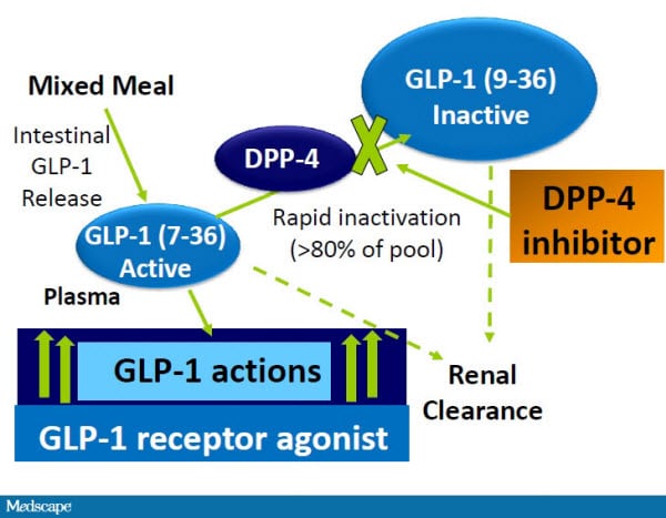
DPP-4 estäjän toimintaperiaate (T2DM)


 http://www.medscape.org/viewarticle/734558

DPP4-estäjät ja GLP-1 analogit

VUODELTA 2005 taustaa DPP4-estäjien kehitelystä


1. 2005. Dipeptidyylipeptidaasi IV estäminen T2DM hoidossa:

Diabetes. 2005 Oct;54(10):2988-94. Dipeptidyl peptidase IV inhibition for the treatment of type 2 diabetes: potential importance of selectivity over dipeptidyl peptidases 8 and 9.Lankas GR1, Leiting B, Roy RS, Eiermann GJ, Beconi MG, Biftu T, Chan CC, Edmondson S, Feeney WP, He H, Ippolito DE, Kim D, Lyons KA, Ok HO, Patel RA, Petrov AN, Pryor KA, Qian X, Reigle L, Woods A, Wu JK, Zaller D, Zhang X, Zhu L, Weber AE, Thornberry NA.
Dipeptidyylipeptidaasi-IV-inhibitio on eräs lähestymistapa 2-tyypin diabeteksen hoidossa. DPP-IV kuuluu seriinipeptidaasien perheeseen ja siihen kuulu lepotilassa olevan solun proliinidipeptidaas (QPP), DPP8 ja DPP9. DPP-IV on inkretiinihormonien avainsäätelijä, mutta muiden perheenjäsenten funktio on ollut  tuntematon.

      • Abstract Dipeptidyl peptidase (DPP)-IV inhibitors are a new approach to the treatment of type 2 diabetes. DPP-IV is a member of a family of serine peptidases that includes quiescent cell proline dipeptidase (QPP), DPP8, and DPP9; DPP-IV is a key regulator of incretin hormones, but the functions of other family members are unknown
Jotta voitiin määrittää selektiivisen DPP-IV inhibition merkitys diabeteksen hoidolle, tehtiin 2 viikon toxisuustutkimuksia ja akuutin toleranssin tutkimuksia koe-eläimellä ja testattiin DPP-IV-, DPP8/DPP9- tai QPP- entsyymien  selektiivisiä estäjiä.
      • To determine the importance of selective DPP-IV inhibition for the treatment of diabetes, we tested selective inhibitors of DPP-IV, DPP8/DPP9, or QPP in 2-week rat toxicity studies and in acute dog tolerability studies.
Koe-eläimessä (rotilla) aiheutui DPP8/DPP9-inhibitiosta alopeciaa, trombosytopeniaa, retikulosytopeniaa, laajentunut perna, histopatologisia monielinmuutoksia ja mortaliteettia. Koirilla DPP8/9-estäjä aiheutti gastrointestinaalista toxisuutta. QPP-estäjä aiheutti retikulosytopeniaa vain rotilla, mutta mitään toxisuutta ei kummallakaan lajilla todettu selektiivisestä DPP-IV estosta.
      • In rats, the DPP8/9 inhibitor produced alopecia, thrombocytopenia, reticulocytopenia, enlarged spleen, multiorgan histopathological changes, and mortality. In dogs, the DPP8/9 inhibitor produced gastrointestinal toxicity. The QPP inhibitor produced reticulocytopenia in rats only, and no toxicities were noted in either species for the selective DPP-IV inhibitor.
Havaittiin myös DPP8/9-estäjän heikentävän T-solujen aktivoitumisen ihmisessä in vitro-mallissa; Näissä mittauksissa selektiivinen DPP-IV-estäjä oli inaktiivisena.
      • The DPP8/9 inhibitor was also shown to attenuate T-cell activation in human in vitro models; a selective DPP-IV inhibitor was inactive in these assays.

Lisäksi tutkijaryhmä havaitsi aiemmin aktiiveiksi immuunifunktiomalleissa raportoitujen DPP-IV-estäjien olevan vahvempia DPP8/9 inhibiittoreina.

Nämä tulokset viittaisivat siihen, että mahdollisten kliinisten kandidaattien selektiivisyyden mittaaminen saattaa olla tärkeää optimaalisen turvallisuusprofiilin takia, koska on kyse tällaisista uuden luokan antihyperglykemisistä vaikuttajista.
      • Moreover, we found DPP-IV inhibitors that were previously reported to be active in models of immune function to be more potent inhibitors of DPP8/9. These results suggest that assessment of selectivity of potential clinical candidates may be important to an optimal safety profile for this new class of antihyperglycemic agents.


Kommentti: NYKYTILANNE. Näitä DPP4-estäjiä on nykyään (2015 jälkeen) käytössä ”inkretiinilääkkeinä”
 
”Det senaste tillskottet i den terapeutiska arsenalen är preparatet som påverkar inkretinsystemet, Glukagonlik peptid 1 (GLP-1- tästä asiasta kirjassa Diabetes typ 2. Läkartidningen Förlag AB 2015 . Sivulta 68.

Tässä kerrotaan miten GLP-1 on glukagonin kaltainen luonnollinen peptidi kehossa ja ehkä tärkein inkretiini, joka vaikuttaa lisäämällä insuliinin erittymistä solunsisäisessä vaiheessa (SU-valmisteiden vaikutuskohdan alapuolelta) ja vain glukoosiarvojen ollessa korkeita.

GLP-1 analogeja on tehty. GLP-1 analogeilla pitkäaikaissokeri laskee 1 %.

Entsyymiestäjä, DPP-4-estäjä taas estää luonnollisen GLP-1-peptidin liian nopean hajoamisen. Suun kautta annettuna tablettina DPP-4 estää entsyymiä dipeptidyylipeptidaasi-4 (DPP4), joka mm. hajoittaa luonnollista GLP1- peptidiä ja GLP-1- vaikutusaikaa kasvaa
Näillä GPP-4-estäjillä saadaan pitkäaikassokeri laskemaan 0,7%

GLP-1 analogeilla on mm. se haitta, että niistä tulee pahoinvointia Ne hidastavat mahan tyhjenemistä ja paino voi hieman laskea.

DPP-4 valmisteet eivät vaikuta painoon. Ne ovat hinnaltaan kalliita Niitten prioriteetti on vasta 10 Sosiaalihallituksen mukaan ja lääkitystä kombinoitaessa vasta kolmannessa vaiheesa tulee tämä valmistekin punnittavaksi. DPP-4 valmistetta voisi ajatella niillä obeeseilla potilailla, joilla olisi tarvetta jostain lisälääkkeestä, mutta jotka saavat hypoglykemioita suun kautta annetusta lisälääkkeestä (sulfonylureavalmisteesta) .

Näiden uusien ja kalliiden läkkeiden kanssa kehoitetaan arvioimaan tehoa 3-6 kuukauden päästä ja lopettamaan lääkitys, jos vaikutusta ei ole tai jos vaikutus on riittämätöntä. (Sivulta 68)

(Nyt 2017 olisi siis voitava odottaa tuloksia siitä, onko DPP4 estäjistä ollut käytännön hyötyä, esim  pitkäaikaissokerin hallintaan ja painon vakautumsieen). 
 
Päivitys 26..9. 2022
 Toistan tämän tekstin  2022 artikkelissa , jossa uusin tulokas  semaglutidi on tullut kysytyksi näissä GLP-1 lääkkeissä etujensa takia. Se on myös painoa laskeva. Ihan niin suosittu Suomessa, että loppuu apteekista syyskuussa 2022. 


tisdag 27 juni 2017

2017 teesi betasolun glutamaattireseptorista KAR

http://eprints.uwe.ac.uk/29385/1/REVISED%20THESIS%20DWOMOH.pdf
Tässä on yli 400 nettisivua ja terminologiasta esitetty  runsas  betasolun tutkimuksissa tarvittu aakkosellinen indeksi
  •  Dwomoh, L. (2017)The role of beta-cell glutamate receptors in pancreatic endocrine function and in the pathogenesis of type 1diabetes mellitus. PhD, University of the West of England. Available from: http://eprints.uwe.ac.uk/29385 We recommend you cite the published version. The publisher’s URL is:
    http://eprints.uwe.ac.uk/29385/
     Background: Kainate receptors (KARs) are one of the three classes of ionotropic glutamate receptors (iGluRs) expressed primarily in the central nervous system (CNS) where they mediate information transfer and neurotransmitter release. Very little is known about native KARs and their interacting partners outside the CNS. Aim: The aim of this study was to investigate systematically the molecular composition and functional properties of KARs in pancreatic endocrine cells and also to investigate the role of KARs in the pathogenesis of T1DM.

Betasolun glutamaattisignalointi: VGLUT

https://en.wikipedia.org/wiki/Glutamate_transporter
Tätä ei ole suomalaisessa wikipediatekstissä, joten otan tähän sitaatin:  Huomaan että tässä artikkelissa  keskitytään aivojen jörejstelmään ja siitä pitää seuloa erikseen se mikä mahtuu pätemään  myös betasolusaarekkeeseen.

SITAATTI:

Glutamate transporters are a family of neurotransmitter transporter proteins that move glutamate – the principal excitatory neurotransmitter – across a membrane. The family of glutamate transporters is composed of two primary subclasses: the excitatory amino acid transporter (EAAT) family and vesicular glutamate transporter (VGLUT) family. In the brain, EAATs remove glutamate from the synaptic cleft and extrasynaptic sites via glutamate reuptake into glial cells and neurons, while VGLUTs move glutamate from the cell cytoplasm into synaptic vesicles. Glutamate transporters also transport aspartate and are present in virtually all peripheral tissues, including the heart, liver, testes, and bone. They exhibit stereoselectivity for L-glutamate but transport both L-aspartate and D-aspartate.
The EAATs are membrane-bound secondary transporters that superficially resemble ion channels.[1] These transporters play the important role of regulating concentrations of glutamate in the extracellular space by transporting it along with other ions across cellular membranes.[2] After glutamate is released as the result of an action potential, glutamate transporters quickly remove it from the extracellular space to keep its levels low, thereby terminating the synaptic transmission.[1][3]
Without the activity of glutamate transporters, glutamate would build up and kill cells in a process called excitotoxicity, in which excessive amounts of glutamate acts as a toxin to neurons by triggering a number of biochemical cascades. The activity of glutamate transporters also allows glutamate to be recycled for repeated releas

Classes

protein gene tissue distribution
EAAT1 SLC1A3 astroglia[5]
EAAT2 SLC1A2 Mainly astroglia;[6] mediates >90% of CNS glutamate reuptake[7]
EAAT3 SLC1A1 all neurons – located on dendrites and axon terminals[8][9]
EAAT4 SLC1A6 neurons
EAAT5 SLC1A7 retina
VGLUT1 SLC17A7 neurons
VGLUT2 SLC17A6 neurons
VGLUT3 SLC17A8 neurons
There are two general classes of glutamate transporters, those that are dependent on an electrochemical gradient of sodium ions (the EAATs) and those that are not (VGLUTs and xCT).[10] The cystine-glutamate antiporter (xCT) is localised to the plasma membrane of cells whilst vesicular glutamate transporters (VGLUTs) are found in the membrane of glutamate-containing synaptic vesicles. Na+-dependent EAATs are also dependent on transmembrane K+ and H+concentration gradients, and so are also known as 'sodium and potassium coupled glutamate transporters'. Na+-dependent transporters have also been called 'high-affinity glutamate transporters', though their glutamate affinity actually varies widely.[10] EAATs are antiporters which carry one molecule of glutamate in along with three Na+ and one H+, while export one K+.[11] EAATs are transmembrane integral proteins which traverse the plasmalemma 8 times.[11]
Mitochondria also possess mechanisms for taking up glutamate that are quite distinct from membrane glutamate transporters.[10]

EAATs

EAAT2 reuptake diagram
This diagram shows the tissue distribution of glutamate transporter 1 (EAAT2) in the brain.[7] EAAT2 is responsible for over 90% of CNS glutamate reuptake.[7][12]
In humans (as well as in rodents), five subtypes have been identified and named EAAT1-5 (SLC1A3, SLC1A2, SLC1A1, SLC1A6, SLC1A7). Subtypes EAAT1-2 are found in membranes of glial cells[13] (astrocytes, microglia, and oligodendrocytes). However, low levels of EAAT2 are also found in the axon-terminals of hippocampal CA3 pyramidal cells.[14] EAAT2 is responsible for over 90% of glutamate reuptake within the central nervous system (CNS).[7][12] The EAAT3-4 subtypes are exclusively neuronal, and are expressed in axon terminals,[8] cell bodies, and dendrites.[9][15] Finally, EAAT5 is only found in the retina where it is principally localized to photoreceptors and bipolar neurons in the retina.[16]
When glutamate is taken up into glial cells by the EAATs, it is converted to glutamine and subsequently transported back into the presynaptic neuron, converted back into glutamate, and taken up into synaptic vesicles by action of the VGLUTs.[3][17] This process is named the glutamate-glutamine cycle.

VGLUTs

Three types of vesicular glutamate transporters are known, VGLUTs 1–3[18] (SLC17A7, SLC17A6, and SLC17A8 respectively)[3] and the novel glutamate/aspartate transporter sialin.[19] These transporters pack the neurotransmitter into synaptic vesicles so that they can be released into the synapse. VGLUTs are dependent on the proton gradient that exists in the secretory system (vesicles being more acidic than the cytosol). VGLUTs have only between one hundredth and one thousandth the affinity for glutamate that EAATs have.[3] Also unlike EAATs, they do not appear to transport aspartate.

VGluT3

VGluT3 (Vesicular Glutamate Transporter 3) that is encoded by the SLC17A8 gene is a member of the vesicular glutamate transporter family that transports glutamate into the cells. It is involved in neurological and pain diseases.
Neurons are able to express VGluT3 when they use a neurotransmitter different to Glutamate, for example in the specific case of central 5-HT neurons.[20][21][22][23] The role of this unconventional transporter (VGluT3) still remains unknown but, at the moment, has been demonstrated that, in auditory system, the VGluT3 is involved in fast excitatory glutamatergic transmission very similar to the another two vesicular glutamate transporter, VGluT1 and VGluT2.[24][25]
There are behavioral and physiological consequences of VGluT3 ablation because it modulates a wide range of neuronal and physiological processes like anxiety, mood regulation, impulsivity, aggressive behavior, pain perception, sleep–wake cycle, appetite, body temperature and sexual behavior. Certainly, no significant change was found in aggression and depression-like behaviors, but in contrast, the loss of VGluT3 resulted in a specific anxiety-related phenotype.
The sensory nerve fibers have different ways to detect the pain hypersensivity throughout their sensory modalities and conduction velocities, but at the moment is still unknown which types of sensory is related to the different forms of inflammatory and neuropathic pain hypersensivity. In this case, Vesicular glutamate transporter 3 (VGluT3), have been implicated in mechanical hypersensitivity after inflammation, but their role in neuropathic pain still remains under debate.
VGluT3 has extensive somatic throughout development, which could be involved in non-synaptic modulation by glutamate in developing retina, and could influence trophic and extra-synaptic neuronal signaling by glutamate in the inner retina.

Pathology

Overactivity of glutamate transporters may result in inadequate synaptic glutamate and may be involved in schizophrenia and other mental illnesses.[1]
During injury processes such as ischemia and traumatic brain injury, the action of glutamate transporters may fail, leading to toxic buildup of glutamate. In fact, their activity may also actually be reversed due to inadequate amounts of adenosine triphosphate to power ATPase pumps, resulting in the loss of the electrochemical ion gradient. Since the direction of glutamate transport depends on the ion gradient, these transporters release glutamate instead of removing it, which results in neurotoxicity due to overactivation of glutamate receptors.[26]
Loss of the Na+-dependent glutamate transporter EAAT2 is suspected to be associated with neurodegenerative diseases such as Alzheimer's disease, Huntington's disease, and ALS–parkinsonism dementia complex.[27] Also, degeneration of motor neurons in the disease amyotrophic lateral sclerosis has been linked to loss of EAAT2 from patients' brains and spinal cords.[27]
Addiction to certain addictive drugs (e.g., cocaine, heroin, alcohol, and nicotine) is correlated with a persistent reduction in the expression of EAAT2 in the nucleus accumbens (NAcc);[28] the reduced expression of EAAT2 in this region is implicated in addictive drug-seeking behavior.[28] In particular, the long-term dysregulation of glutamate neurotransmission in the NAcc of addicts is associated with an increase in vulnerability to relapse after re-exposure to the addictive drug or its associated drug cues.[28] Drugs which help to normalize the expression of EAAT2 in this region, such as N-acetylcysteine, have been proposed as an adjunct therapy for the treatment of addiction to cocaine, nicotine, alcohol, and other drugs.[28]

See also

måndag 26 juni 2017

Betasolun glutamaattissignalointi. NMDAR. VGLUT

 Haimasaarekkeen  mielenkiintoiset ajat: Glutamaattisignalointi endokriinisissä soluissa 

(Kommentti:  Artikkeliin liittyy kuva, jossa on solun sisäisen insuliinijyväsen pintaan liitetty VGLUT niminen proteiini. Siitä erikseen)

 LÄHDE:
https://www.ncbi.nlm.nih.gov/pubmed/26740469
 trends Endocrinol Metab. 2016 Mar;27(3):177-88. doi: 10.1016/j.tem.2015.12.004. Epub 2015 Dec 28. Exciting Times for Pancreatic Islets: Glutamate Signaling in Endocrine Cells.

Tiivistelmän suomennosta, Abstract

 Glutamaatti eli glutamiinihappo (Glu lyhennys, koodi E)  on keskushermoston excitatorinen hermonvälittäjäaine.Se myös moduloi  haimasaarekkeen endokriinisten solujen funktiota ja elinkykyisyyttä.  Insuliinia erittävissä betasoluissa glutamaatti toimii solunsisäisenä lähettinä ja sen kuljetus sekretorisiin jyväsiin edistää GSIS , glukoosin stimuloimaa ja inkretiinin stimuloimaa insuliinin eritystä.  Mitokondriassa tapahtuva glutamaatin hajoaminen osallistuu myös insuliinin vapautumiseen, kun glutamaattidehydrogenaasi aktivoituu allosteerisesti.
Glutamaatti voi antaa myös extrasellulaarista signaalia glutamaattireseptorien (AMPA, NMDA) kautta ja moduloida glukagonim, insuliinin ja somatostatiinin eritystä sekä saarekesolun elossapysymistä . Hajoamistuotteet  GABA ja gamma-OH-voihappo vapautuvat ja vaikutatvat myös betasolun käyttäytymiseen.  Täten  saarekkeen glutamattireseptorit, kuten NMDA-reseptorit, saataisivat toimia mahdollisina  lääkkeen kohteina- kun ollaan kehiteelemässä uusia lääkkeitä diabeteksen lisäterapiaksi.
  • Glutamate represents a key excitatory neurotransmitter in the central nervous system, and also modulates the function and viability of endocrine cells in pancreatic islets. In insulin-secreting beta cells, glutamate acts as an intracellular messenger, and its transport into secretory granules promotes glucose- and incretin-stimulated insulin secretion. Mitochondrial degradation of glutamate also contributes to insulin release when glutamate dehydrogenase is allosterically activated. It also signals extracellularly via glutamate receptors (AMPA and NMDA receptors) to modulate glucagon, insulin and somatostatin secretion, and islet cell survival. Its degradation products, GABA and γ-hydroxybutyrate, are released and also influence islet cell behavior. Thus, islet glutamate receptors, such as the NMDA receptors, might serve as possible drug targets to develop new medications for adjunct treatment of diabetes.
 Bildresultat för islet cell AMPA and GLu signalling


Nykyisistä trendeistä        Trends

 Tyypin 2 diabeteksella ja tavallisilla neurodegeneratiivisilla taudeilla  on samoja patologisia mekanismeja kuten excitotoksinen  solukuolema.
Keskushermoston pääasiallisin exkitatorinen  hermonvälittäjäaine glutamaatti  omaa myös tärkeän osan haimasaarekkeen toiminnassa ja saarekkeen elossapysymisessä.
  • Type 2 diabetes and common neurodegenerative diseases share similar pathomechanisms, including excitotoxic cell death.
  • Glutamate, the major excitatory neurotransmitter in the central nervous system, plays an important role in islet cell function and islet cell survival.
 NMDA-reseptori on glutamaattireseptoreita . Sen vastavaikuttajilla NMDA-antagonisteilla, on verensokeria madaltavia  ja seerumin insuliinipitoisuuksia  lisääviä vaikutuksia  pääasiassa sokerirasitustestin (OGT)  ensimmäisessä vaiheessa niillä  yksilöillä, joilla on  T2DM
  • NMDARs are glutamate receptors that may be potential drug targets for the treatment of type 2 diabetes.
 Dextrometrofaanilla on insuliinin säätelyssä mm. NMDAR antagonistivaikutusta ja sillä  on   veren sokeria laskevia vaikutuksia ja se lisää seerumin insuliinin pitoisuuksia  pääasiassa OGT:n ensimmäisessä vaiheessa  yksilöillä joilla on T2DM
  • Dextromethorphan, an over-the-counter drug and antagonist of NMDARs, has blood glucose lowering effects and increases serum insulin concentrations mainly during the first phase of an oral glucose tolerance test in individuals with type 2 diabetes.


Bildresultat för islet cell AMPA and GLu signalling