Etiketter

Summa sidvisningar

Leta i den här bloggen

tisdag 27 november 2018

OGT, O-GlcNAcylaatio ja ubikitinaatiojärjestelmä

17. Ruan H-B, Nie Y, Yang X. Regulation of protein degradation by O-GlcNAcylation: crosstalk with ubiquitination. Mol Cell Proteomics (2013) 12:3489–97. doi: 10.1074/mcp.R113.029751
PubMed Abstract | CrossRef Full Text | Google Scholar

måndag 26 november 2018

Lihas

 https://www.ncbi.nlm.nih.gov/pubmed/30459708

https://www.frontiersin.org/files/Articles/409446/fendo-09-00578-HTML/image_m/fendo-09-00578-g001.jpg

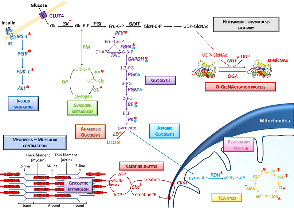
 Edustava kaava luustolihaksessa tapahtuvasta heksosaminibiosynteesitiestä,  jossa muodostuu O-GlcNAc  ja tapahtuu posttranslationaalinen lisääminen   solussa oleviin proteiineihin  transferaasilla OGT / tai  tämän ryhmän poisto  OGA hydrolaasilla). kuvan oiekassa yläkulmassa.
kuvassa esitetään myös  miten insuliini signaloituminen tapahtuu IR insuliinireseptoriteitse, jolloin aktivoituu signaalitie IRS-1- PI3K-PDK1-Akt.
Glukoosin tulo soluun tapahtuu GLUT4-proteiinin avulla ja  sitten kinaasi fosforyloi  sen Glukoosi-6 fosfaatiksi.  Siitä voi osa mennä glykogeeniaineenvaihduntaan, jolloin sen täytyy ensin muuttau Glukoosi-1-fosfaatiksi ja aktivoitua UTP-energialla.  UDP-glukoosiksi, josta voi syntetisoida  lihasglykogeeniä.
Osa soluun tulleesta glukoosista jatkaa muuttumalla fruktoosiksi Fru-6-P ja siten  fruktoosi-bifosfaatiksi ja  menee glykolyysiteihen.  Palorypälehappovaiheesta  voi osa mennä  anaaerobiseen glykolyysiin  tarvittaessa ja siitä tulee vähän ATP:tä ja lopulta  maitohappoa.  Aerobinen glykolyysi tuottaa paljon ATP:tä mitokondriasta. Lihaksessa ATP-energiaa voidaan  slussata  lihakseen  kreatiniinislussin avulla . Myosiinissa on omia ATPaaseja.  Entsyymien nimiå alla. Ne kuuluvat kuvan selitykseen.

  • Representative scheme of the presence of O-GlcNAcylation on glucose metabolism in skeletal muscle. Several signaling and metabolic pathways are indicated, in particular the hexosamine biosynthesis pathway, glycolysis (anaerobic and aerobic glycolysis), glycogen metabolism, insulin signaling, and TCA cycle. Specific molecular components of skeletal muscle such as myofibrils and creatine shuttle are also represented. Red asterisks correspond to O-GlcNAcylated proteins; blue asterisks correspond to enzymes including in protein-protein complexes (such as the glycolytic metabolon) which could be potentially modulated consecutively to O-GlcNAcylation changes in skeletal muscle cells. A, Aconitase; BE, Beta-enolase; CKc, Creatine kinase cytoplasmic; CKm, Creatine kinase mitochondrial; CS, Citrate synthase; FBPA, Fructose-bisphosphate aldolase; FH, Fumarate hydratase; GAPDH, Glyceraldehyde-3-phosphate dehydrogenase; GFAT, Glutamine,fructose-6-phosphate aminotransferase; GK, Glucokinase; GP, Glycogen phosphorylase; GS, Glycogen synthase; IDH, Isocitrate dehydrogenase; KGD, Ketoglutarate dehydrogenase; LD, Lactate dehydrogenase; MDH, Malate dehydrogenase; OGA, O-GlcNAcase; OGT, O-GlcNAc transferase; PDH, Pyruvate Dehydrogenase; PFK, Phosphofructokinase; PGI, Phosphoglucose isomerase; PGK, Phosphoglycerate kinase; PGM, Phosphoglycerate mutase; PK, Pyruvate kinase; PM, phosphoglucomutase; PP, UDP-glucose pyrophosphorylase; SDH, Succinate Dehydrogenase; SL, Succinyl-CoA ligase; TPI, Triose-phosphate isomerase.
 Abstraktista suomennosta:
LUUSTOLIHAS  ( tahdonalainen lihaksisto)  käsittää 40 % koko kehonpainosta. Lihaksen päätehtävä on konvertoida kemiallinen energia mekaaniseksi energiaksi, mikä  takaa  voiman generoitumista  liikkeisiin ja lokomotioon ja pitää yllä asentoa. Ratkaisevasta   energiariippuvuudesta  vastaa lihaksisto, joka on keskeinen koko kehon  aminohappojen ja hiilihdydraattien varasto. Juuri näistä ravintoaineista muun muuassa riippuu posttranslationaalinen modifikaatio  N-asetyyliglukosaminin  (GlucNAc)   liittäminen  -O- asemaan  entsyymeissä (O-GlucNAc-) Siinä siirretään tai tarvitatessa  irrotetaan näitä ainutlaatuisia monosakkarideja ( N-asetyyli-D-glukosamineja)  aminohappojen seriinin tai treoniini OH-ryhmään  nukleosytoplasmisessa tai mitokondriaalisessa proteiinissa. Prosessi on dynaaminen: ja tapahtuu entsyymien O-GlcNAc- transferaasin (OGT) tai vastaavasti  O-GlcNAc-glykosidihydrolaasin (OGA) katalysoimina reaktioina. Tämä  heksosamini on rakenteellinen sokeri ja sillä on paljon merkitystä typpipitoisena  aminosokerina solujen pintarakenteissa ja proteiinien ikää pidentävissä tekijöissä.

OGT = O-linkkiytynyt N-asetyyliglukosaminyylitransferaasi
OGA , 3-O-(N-asetyyli-D-glukosaminyyli) L-seriini/treoniini N-asetyyliglukosaminyylihydrolaasi
tai lyhemmin sanottuna    O-GlcNAcaasi

(Selitys: 
Glukosamini on  hiilihydraattiaineenvaihdunnasta peräisin oleva sokeri   glukoosi, jossa on  aminohapon glutamiinihapon amidityppi (N)  liitetty sokeriin.
Tämä glukosamini on sitten asetyloitu, saanut Ac- ryhmänsä.
Nyt se on GlcNAc
Sitten se on liitetty  proteiinissa olevan  aminohapon OH- ryhmään sen O -molekyyliin, joten saadaan O-GlcNAc . Nyt on sokeriaineenvaihdunnasta kehittynyt rakenteellinen molekyyli, jonka avulla   saadaan erilaisia glykokonjugaatteja  proteiineille. Ihmiskehon rakenneaminohapoista  seriinillä ja treoniinilla on OH- ryhmät rakenteessa.   Seriini ja treoniini (s ja t) kuuluvat niihin aminohappoihin joita ihmisen genomi koodaa).

  • Abstract

  • Skeletal muscle represents around 40% of whole body mass. The principal function of skeletal muscle is the conversion of chemical energy toward mechanic energy to ensure the development of force, provide movement and locomotion, and maintain posture. This crucial energy dependence is maintained by the faculty of the skeletal muscle for being a central place as a "reservoir" of amino acids and carbohydrates in the whole body. A fundamental post-translational modification, named O-GlcNAcylation, depends, inter alia, on these nutrients; it consists to the transfer or the removal of a unique monosaccharide (N-acetyl-D-glucosamine) to a serine or threonine hydroxyl group of nucleocytoplasmic and mitochondrial proteins in a dynamic process by the O-GlcNAc Transferase (OGT) and the O-GlcNAcase (OGA), respectively. 
O-GlucNAc- rakenteen liittämisen  on havaittu  osallistuvan vahvasti  solunsisäiseen aineenvaihduntaan  eri elimissä ja kudoksissa, sollä se moduloi  signalointiteitä, geeni-ilmentymää ja sytoskeletaalisia funktioita.  Tällaista on havaittu aivosta, maksasta, munuaisesta ja haimasta ja   O-GlcNAc  linkkiytyy  eri tautien etiologiaankin.   Viime vuosina on monet tutkimukset kohdistuneet O-GlucNAc- liitännän  osuuteen luustolihaksen fysiologiassa ja patofysiologiassa. Näissä tutkimuksissa on päämielenkiinto kohdistunut  O-GlcNAc-liittymiin lihastreenin aikana tai lihaksen kuihtumistiloissa.  Suurin osa tutkimuksista viittaa  siihen, että  esiintyy erilaista O-GlcNAc-liittymäleimaa riippuen lihakselle tyypillisestä  aineenvaihdunnasta levossa, lihaksen kuihtumissa  tai treenausolosuhteissa, samoin riippuen   treenausohjelmasta, siitä onko kyse  äkkinäsiestä  vai pitkäaikaisesta  voimanponnistuksesta.
 Ensimmäiset saadut oivallukset  antoivat viitettä   jonkin verran erilaisiin OGT/OGA ilmentymisiin ja /tai aktiivisuuksiin  ja erilaisiin solustressivasteisiin (ROS, reaktiivisiin happilajeihin ja/tai  HSP-proteiineihin).
Vankkaa tietoa  tuli  siitä, että  nämä O-GluNAc-muutokset  voisivat johtaa
(1)  hiilihydraattiaineenvaihdunnan erilaisiin moduloitumisiin, koska tiedetään  suurimman osan entsyymeistä  saavan  posttranslationaalisen  O-GlcNAc- lisukkeen ja
 (2) proteiinisynteesi/proteiininhajoitustasapainon erilaiseen modulaatioon, koska  O-GlcNAc -päätteen saaminen säätelee luustolihaksessa  muutamia avainasemassa olevia signaaliteitä kuten  Akt/GSK3beeta, Akt/mTOR, myogeniini/atrogiini-1, myogeniini/Mef2D, Mrf4 ja PGC-1alfa.

  • O-GlcNAcylation has been shown to be strongly involved in crucial intracellular mechanisms through the modulation of signaling pathways, gene expression, or cytoskeletal functions in various organs and tissues, such as the brain, liver, kidney or pancreas, and linked to the etiology of associated diseases. In recent years, several studies were also focused on the role of O-GlcNAcylation in the physiology and the physiopathology of skeletal muscle. These studies were mostly interested in O-GlcNAcylation during muscle exercise or muscle-wasting conditions. Major findings pointed out a different "O-GlcNAc signature" depending on muscle type metabolism at resting, wasting and exercise conditions, as well as depending on acute or long-term exhausting exercise protocol. First insights showed some differential OGT/OGA expression and/or activity associated with some differential stress cellular responses through Reactive Oxygen Species and/or Heat-Shock Proteins. Robust data displayed that these O-GlcNAc changes could lead to (i) a differential modulation of the carbohydrates metabolism, since the majority of enzymes are known to be O-GlcNAcylated, and to (ii) a differential modulation of the protein synthesis/degradation balance since O-GlcNAcylation regulates some key signaling pathways such as Akt/GSK3β, Akt/mTOR, Myogenin/Atrogin-1, Myogenin/Mef2D, Mrf4 and PGC-1α in the skeletal muscle. 
 Lopuksi O-GlcNAc -siirron osallistuminen  joihinkin luustolihaksen metabolisiin prosesseihin saattaisi linkkiytyä  joihinkin liitännäistauteihin kuten tyypin 2 diabetekseen tai neuromuskulaarisiin sairauksiin, joissa  näyttäa olevan yleisesti kriittisesti kohonnutta  O-GlcNAc liitäntää.
  • Finally, such involvement of O-GlcNAcylation in some metabolic processes of the skeletal muscle might be linked to some associated diseases such as type 2 diabetes or neuromuscular diseases showing a critical increase of the global O-GlcNAcylation level.

E3 ubikitiiniligaasi sokeriaineenaihdunnasta

 F(1,6) bifosfataasin entsymin hajoittaa  u´E3 ubikitiiniligaasi, joka hajoaa myös itse proteosomilla.


Muut E3 ubikitiiniligaasit
  •  HiilihydraattiaineenvaihduntaHiivassa:
Mol Biol Cell. 2008 Aug; 19(8): 3323–3333.
PMCID: PMC2488282
PMID: 18508925
The Yeast GID Complex, a Novel Ubiquitin Ligase (E3) Involved in the Regulation of Carbohydrate Metabolism

Thomas Sommer, Monitoring Editor

The Gid complex, a novel ubiquitin ligase (E3) required for the degradation of the key gluconeogenic enzyme fructose-1,6-bisphosphatase. The Gid complex binds to FBPase, when S. cerevisiae cells are growing on an ethanol-containing medium. On shift of cells to glucose, Gid4/Vid24 occurs and activates the complex, which then polyubiquitinates FBPase before its degradation by the proteasome. Gid4/Vid24 is itself degraded by the proteasome.

PEPCK, another gluconeogenic enzyme, is also subject to catabolite degradation (Holzer, 1976 ; Muller et al., 1981 ). As Figure 7 shows, it is stabilized in GID2/RMD5 and GID4/VID24 deleted cells. Thus, Gid complex-dependent degradation is not restricted to FBPase but plays a more general role within the regulation of carbohydrate metabolism.
Gid protein homologues were also found to form a complex in mammals (Kobayashi et al., 2007 ). Although no function for this CTLH complex has been described, one subunit has been implicated in proteasomal degradation of α-catenin (Suzuki et al., 2008 ). This suggests that the CTLH complex, like the Gid complex, might also bear E3 activity.
In conclusion (Figure 8), our study shows that the Gid complex is a new ubiquitin ligase with novel types of subunits involved in catabolite degradation of gluconeogenic enzymes in yeast. We also identify Vid24/Gid4 as an important regulator of its ubiquitin ligase activity.

ENTÄ tämän E3 ubikitiiniligaasihomologin  tehtävät ihmisessä?

Löytyy kaksi geeniä
RMD5A, GID2A, CTLH , Kr. 2 required for meiotic division
RMD5B, GID2B, kr.5
required ofr meiotic nuclear division
Katson näistä  enemmän tietoa  onko niillä  yhteyttä sokeriaineenvaihduntaan kuten hiivassa, josa se hajoitaa fruktoosi1,6,-bifosfaataasia.


Fruktoosi-1,6-bifosfataasi

https://sv.wikipedia.org/wiki/Fruktos-1,6-bisfosfatas

Fruktos-1,6-bisfosfatas

Fruktos-1,6-bisfosfatas-oktamer, sulfolobus tokodaii.
Fruktos-1,6-bisfosfatas är ett enzym som ingår i glukoneogenesen och därmed också i coricykeln. Det katalyserar defosforyleringen av fruktos-1,6-bisfosfat till fruktos-6-fosfat. Det aktiveras av höga nivåer av ATP i sin omgivande miljö, men utnyttjar inte något ATP för att kunna drivas. Enzymet arbetar vid motsatt intracellulär miljö än dess motsvarande enzym i glykolysen, fosfofruktokinas.

 Suomi:
 Fruktoosi-1,6-bisfosfataasi on entsyymi, joka katalysoi fruktoosi-1,6-bisfosfaatin hydrolyysiä fruktoosi-6-fosfaatiksi glukoneogeneesissä. Entsyymi on tärkeä glukoneogeneesin säätelykohta.[1] Fruktoosi-1,6-bisfosfataasin EC-numero on EC 3.1.3.11.[2] Eläimillä fruktoosi-1,6-bisfosfataasia esiintyy maksassa ja munuaisissa
  1. Ihmisen fruktoosi-1,6-bisfosfataasi on rakenteeltaan tetrameeri. Eräillä kasveilla, kuten herneellä se on monomeeri ja eräillä bakteereilla, kuten Escherichia colilla se on rakenteeltaan monomeeri. Toimiakseen entsyymi tarvitsee koentsyymikseen Mg2+-ionin tai Mn2+-ionin.[2]
    Fruktoosi-1,6-bisfosfataasin katalysoiman reaktion mekanismista ei ole täyttä varmuutta ja esitettyjä mekanismeja on kaksi: dissosiatiivinen ja assosiatiivinen mekanismi. Assosiatiivisessa mekanismissa vesimolekyyli hyökkää entsyymin katalysoimana fruktoosi-1,6-bisfosfaatin 1-asemassa sijaitsevan fosfaattiryhmän fosforiatomiin ja fosfaattiryhmä poistuu vetyfosfaatti-ionina ja entsyymin aktiivisen keskuksen asparagiinihappo luovuttaa protonin, jolloin muodostuu hydroksyyliryhmä ja fruktoosi-6-fosfaatti vapautuu. Dissosiatiivisessa mekanismissa hyökkäys tapahtuu fosfaattiesteriryhmän happeen, jolloin alkoholiryhmä vapautuu ja seuraavassa vaiheessa fosfaatti-ionista muodostuu vetyfosfaatti. Myös tässä mekanismissa asparagiinihappo on tärkeässä osassa. Krystallografiset tutkimukset tukevat kumpaakin mekanismia.[3]
    Kun solujen energiavarastot ovat vähäiset, on soluissa paljon AMP:tä, joka on fruktoosi-1,6-fosfataasin inhibiittori. Tällöin entsyymin toiminta estyy.
    ATP sen sijaan aktivoi fruktoosi-1,6-bisfosfataasia.
     Fruktoosi-2,6-bisfosfaatti on voimakas entsyymin allosteerinen inhibiittori. Fruktoosi-2,6-bisfosfaatin määrä riippuu veren glukagonipitoisuudesta.
    ATP:n lisäksi myös sitraatti-ioni aktivoi fruktoosi-1,6-bisfosfataasia.[1][4]
    Fruktoosi-1,6-bisfosfataasin puutos on harvinainen autosomien välityksellä resessiivisesti periytyvä sairaus.
     Entsyymin puutos aiheuttaa laktista asidoosia ja hypoglykemiaa. Pienillä lapsina tämä ilmenee muun muassa hyperventiloimisena, ärtyisyytenä, unettomuutena ja lihasten heikkoutena. Hoitona on suun kautta tai suonensisäisesti annettava glukoosi ja asidoosin hoitoon vetykarbonaattiliuos.[5]
    Lähteet
  2. Jeremy M. Berg, John L. Tymoczko & Lubert Stryer: Biochemistry, 6th Edition, s. 461, 466. W. H. Freeman and Company, 2006. ISBN 978-0-7167-8724-2. (englanniksi)
  3. EC 3.1.3.11 - fructose-bisphosphatase Brenda. Viitattu 29.08.2013. (englanniksi)
  4. John McMurry, Tadhg P. Begley: The organic chemistry of biological pathways, s. 20. Roberts and Company Publishers, 2005. ISBN 978-0974707716. Kirja Googlen teoshaussa (viitattu 29.08.2013). (englanniksi)
  5. Richard A Harvey,Denise R Ferrier: Lippincott's Illustrated Reviews: Biochemistry, s. 121. Lippincott Williams & Wilkins, 2010. ISBN 978-1-60831-412-6. Kirja Googlen teoshaussa. (englanniksi)
  6. John Fernandes: Inborn metabolic diseases: diagnosis and treatment, s. 528. Springer, 2006. ISBN 978-3540287834. Kirja Googlen teoshaussa (viitattu 29.08.2013). (englanniksi)
.

RMD5B, GID2B (5q35.3) E3 ubikitiiniligaasi, tärkeä tuman meioottiselle jaolle.

https://www.ncbi.nlm.nih.gov/gene
Preferred Names
E3 ubiquitin-protein transferase RMND5B; protein RMD5 homolog B
Names
GID complex subunit 2 homolog B

Related articles in PubMed

RMND5A, GID2A,Geenin normaalius on meiossissa ja hermostonkehitykselle tärkeä.

https://www.ncbi.nlm.nih.gov/gene/64795
Also known as
CTLH; GID2; RMD5; GID2A; p44CTLH
Expression
Ubiquitous expression in esophagus (RPKM 17.5), heart (RPKM 16.4) and 25 other tissues See more
Orthologs mouse
Preferred Names
E3 ubiquitin-protein transferase RMND5A
Names
44-kD protein coding for CTLH motif
C-terminal to LisH motif, 44 kDa
GID complex subunit 2 homolog A
protein RMD5 homolog A
NP_073617.1

Related articles in PubMed

  1. NM_022780.4NP_073617.1  E3 ubiquitin-protein transferase RMND5A
    See identical proteins and their annotated locations for NP_073617.1
    Status: VALIDATED
    Source sequence(s)
    AC064848, AK023972, BC012165, BC047668, BU620245
    Consensus CDS
    CCDS1991.1
    UniProtKB/Swiss-Prot
    Q9H871
    Related
    ENSP00000283632.4, OTTHUMP00000160759, ENST00000283632.4
    Conserved Domains (3) summary
    smart00667
    Location:114146
    LisH; Lissencephaly type-1-like homology motif
    pfam10607
    Location:153297
    CLTH; CTLH/CRA C-terminal to LisH motif domain
    cd16794
    Location:333381
    dRING_RMD5A; Degenerated RING finger found in protein RMD5 homolo

E3 ubiquitin-protein transferase RMND5A [Homo sapiens]

NCBI Reference Sequence: NP_073617.1
LOCUS       NP_073617                391 aa            linear   PRI 23-NOV-2018
DEFINITION  E3 ubiquitin-protein transferase RMND5A [Homo sapiens].
ACCESSION   NP_073617
VERSION     NP_073617.1
DBSOURCE    REFSEQ: accession NM_022780.4
KEYWORDS    RefSeq.
SOURCE      Homo sapiens (human)
  ORGANISM  Homo sapiens
            Eukaryota; Metazoa; Chordata; Craniata; Vertebrata; Euteleostomi;
            Mammalia; Eutheria; Euarchontoglires; Primates; Haplorrhini;
            Catarrhini; Hominidae; Homo.
REFERENCE   1  (residues 1 to 391)
  AUTHORS   Lampert F, Stafa D, Goga A, Soste MV, Gilberto S, Olieric N,
            Picotti P, Stoffel M and Peter M.
  TITLE     The multi-subunit GID/CTLH E3 ubiquitin ligase promotes cell
            proliferation and targets the transcription factor Hbp1 for
            degradation
  JOURNAL   Elife 7, e35528 (2018)
   PUBMED   29911972
  REMARK    Publication Status: Online-Only
REFERENCE   2  (residues 1 to 391)
  AUTHORS   Boldt K, van Reeuwijk J, Lu Q, Koutroumpas K, Nguyen TM, Texier Y,
            van Beersum SE, Horn N, Willer JR, Mans DA, Dougherty G, Lamers IJ,
            Coene KL, Arts HH, Betts MJ, Beyer T, Bolat E, Gloeckner CJ,
            Haidari K, Hetterschijt L, Iaconis D, Jenkins D, Klose F, Knapp B,
            Latour B, Letteboer SJ, Marcelis CL, Mitic D, Morleo M, Oud MM,
            Riemersma M, Rix S, Terhal PA, Toedt G, van Dam TJ, de Vrieze E,
            Wissinger Y, Wu KM, Apic G, Beales PL, Blacque OE, Gibson TJ,
            Huynen MA, Katsanis N, Kremer H, Omran H, van Wijk E, Wolfrum U,
            Kepes F, Davis EE, Franco B, Giles RH, Ueffing M, Russell RB and
            Roepman R.
  CONSRTM   UK10K Rare Diseases Group
  TITLE     An organelle-specific protein landscape identifies novel diseases
            and molecular mechanisms
  JOURNAL   Nat Commun 7, 11491 (2016)
   PUBMED   27173435
  REMARK    Publication Status: Online-Only
REFERENCE   3  (residues 1 to 391)
  AUTHORS   Li J, Chen Y, Qin X, Wen J, Ding H, Xia W, Li S, Su X, Wang W, Li
            H, Zhao Q, Fang T, Qu L and Shao N.
  TITLE     MiR-138 downregulates miRNA processing in HeLa cells by targeting
            RMND5A and decreasing Exportin-5 stability
  JOURNAL   Nucleic Acids Res. 42 (1), 458-474 (2014)
   PUBMED   24057215
  REMARK    GeneRIF: The study identifies a miR-138-RMND5A-Exportin-5 as a
            previously unknown miRNA processing regulatory pathway in HeLa
            cells.
REFERENCE   4  (residues 1 to 391)
  AUTHORS   Francis O, Han F and Adams JC.
  TITLE     Molecular phylogeny of a RING E3 ubiquitin ligase, conserved in
            eukaryotic cells and dominated by homologous components, the
            muskelin/RanBPM/CTLH complex
  JOURNAL   PLoS ONE 8 (10), e75217 (2013)
   PUBMED   24143168
  REMARK    Erratum:[PLoS One. 2013 Nov 8;8(11):null. PMID: 29161723]
            Publication Status: Online-Only
REFERENCE   5  (residues 1 to 391)
  AUTHORS   Vogel TW, Manjila S and Cohen AR.
  TITLE     Novel neurodevelopmental disorder in the case of a giant
            occipitoparietal meningoencephalocele
  JOURNAL   J Neurosurg Pediatr 10 (1), 25-29 (2012)
   PUBMED   22681319
  REMARK    GeneRIF: Duplications of this region involving RMND5A, whose
            product contains a C-terminal to lis homology (LisH) domain, have
            not previously been associated with a defined phenotype but may
            present insight into encephalocele formation.
REFERENCE   6  (residues 1 to 391)
  AUTHORS   Kobayashi N, Yang J, Ueda A, Suzuki T, Tomaru K, Takeno M, Okuda K
            and Ishigatsubo Y.
  TITLE     RanBPM, Muskelin, p48EMLP, p44CTLH, and the armadillo-repeat
            proteins ARMC8alpha and ARMC8beta are components of the CTLH
            complex
  JOURNAL   Gene 396 (2), 236-247 (2007)
   PUBMED   17467196
  REMARK    GeneRIF: RanBPM, ARMC8alpha, ARMC8beta, Muskelin, p48EMLP, and
            p44CTLH form complexes in cells.
REFERENCE   7  (residues 1 to 391)
  AUTHORS   Bowzard JB, Cheng D, Peng J and Kahn RA.
  TITLE     ELMOD2 is an Arl2 GTPase-activating protein that also acts on Arfs
  JOURNAL   J. Biol. Chem. 282 (24), 17568-17580 (2007)
   PUBMED   17452337
REFERENCE   8  (residues 1 to 391)
  AUTHORS   Colland F, Jacq X, Trouplin V, Mougin C, Groizeleau C, Hamburger A,
            Meil A, Wojcik J, Legrain P and Gauthier JM.
  TITLE     Functional proteomics mapping of a human signaling pathway
  JOURNAL   Genome Res. 14 (7), 1324-1332 (2004)
   PUBMED   15231748
COMMENT     VALIDATED REFSEQ: This record has undergone validation or
            preliminary review. The reference sequence was derived from
            AK023972.1, BC047668.1, BC012165.1, AC064848.5 and BU620245.1.
            
            Sequence Note: The RefSeq transcript and protein were derived from
            transcript and genomic sequence to make the sequence consistent
            with the reference genome assembly. The genomic coordinates used
            for the transcript record were based on alignments.
            
            ##Evidence-Data-START##
            Transcript exon combination :: SRR1803616.90910.1,
                                           SRR1660805.158266.1 [ECO:0000332]
            RNAseq introns              :: single sample supports all introns
                                           SAMEA1965299, SAMEA1966682
                                           [ECO:0000348]
            ##Evidence-Data-END##
FEATURES             Location/Qualifiers
     source          1..391
                     /organism="Homo sapiens"
                     /db_xref="taxon:9606"
                     /chromosome="2"
                     /map="2p11.2"
     Protein         1..391
                     /product="E3 ubiquitin-protein transferase RMND5A"
                     /EC_number="2.3.2.27"
                     /note="C-terminal to LisH motif, 44 kDa; protein RMD5
                     homolog A; GID complex subunit 2 homolog A; 44-kD protein
                     coding for CTLH motif"
                     /calculated_mol_wt=43862
     Site            1
                     /site_type="other"
                     /experiment="experimental evidence, no additional details
                     recorded"
                     /note="N-acetylmethionine. {ECO:0000244|PubMed:22814378};
                     propagated from UniProtKB/Swiss-Prot (Q9H871.1)"
     Region          114..146
                     /region_name="LisH"
                     /note="Lissencephaly type-1-like homology motif;
                     smart00667"
                     /db_xref="CDD:128913"
     Region          153..297
                     /region_name="CLTH"
                     /note="CTLH/CRA C-terminal to LisH motif domain;
                     pfam10607"
                     /db_xref="CDD:313761"
     Region          333..381
                     /region_name="dRING_RMD5A"
                     /note="Degenerated RING finger found in protein RMD5
                     homolog A (RMD5A); cd16794"
                     /db_xref="CDD:319708"
     Region          336..377
                     /region_name="degenerated RING finger"
                     /note="degenerated RING finger [structural motif]"
                     /db_xref="CDD:319708"
     CDS             1..391
                     /gene="RMND5A"
                     /gene_synonym="CTLH; GID2; GID2A; p44CTLH; RMD5"
                     /coded_by="NM_022780.4:378..1553"
                     /db_xref="CCDS:CCDS1991.1"
                     /db_xref="GeneID:64795"
                     /db_xref="HGNC:HGNC:25850"
ORIGIN      
        1 mdqcvtvere lekvlhkfsg ygqlcergle elidytgglk heilqshgqd aelsgtlslv
       61 ltqcckrikd tvqklasdhk dihssvsrvg kaidknfdsd issvgidgcw qadsqrllne
      121 vmvehffrqg mldvaeelcq esglsvdpsq kepfvelnri lealkvrvlr palewavsnr
      181 emliaqnssl efklhrlyfi sllmggttnq realqyaknf qpfalnhqkd iqvlmgslvy
      241 lrqgienspy vhlldanqwa dicdiftrda callglsves plsvsfsagc valpalinik
      301 avieqrqctg vwnqkdelpi evdlgkkcwy hsifacpilr qqttdnnppm klvcghiisr
      361 dalnkmfngs klkcpycpme qspgdakqif f
//
  1. (RMD5A)

GNPDA1 (5q31.3), oskilliini, glcN6P deaminaasi 1

https://www.ncbi.nlm.nih.gov/gene/10007

GPI; HLN; GNP1; GNPI; GNPDA
Summary
Glucosamine-6-phosphate deaminase (EC 3.5.99.6) is an allosteric enzyme that catalyzes the reversible conversion of D-glucosamine-6-phosphate into D-fructose-6-phosphate and ammonium (Arreola et al., 2003 [PubMed 12965206]).[supplied by OMIM, Jan 2010]

Preferred Names
glucosamine-6-phosphate isomerase 1
Names
GNPDA 1
glcN6P deaminase 1
oscillin

Related articles in PubMed

PubMed

GeneRIFs: Gene References Into FunctionsWhat's a GeneRIF?

peptidi rakenne, historia, struktuuri
Konservoitu domeeni
SugarP_isomerase: Sugar Phosphate Isomerase family; includes type A ribose 5-phosphate isomerase (RPI_A), glucosamine-6-phosphate (GlcN6P) deaminase, and 6-phosphogluconolactonase (6PGL). RPI catalyzes the reversible conversion of ribose-5-phosphate to ribulose 5-phosphate, the first step of the non-oxidative branch of the pentose phosphate pathway. GlcN6P deaminase catalyzes the reversible conversion of GlcN6P to D-fructose-6-phosphate (Fru6P) and ammonium, the last step of the metabolic pathway of N-acetyl-D-glucosamine-6-phosphate. 6PGL converts 6-phosphoglucono-1,5-lactone to 6-phosphogluconate, the second step of the oxidative phase of the pentose phosphate pathway.

glucosamine-6-phosphate isomerase 1 [Homo sapiens]

NCBI Reference Sequence: NP_005462.1
LOCUS       NP_005462                289 aa            linear   PRI 22-NOV-2018
DEFINITION  glucosamine-6-phosphate isomerase 1 [Homo sapiens].
ACCESSION   NP_005462
VERSION     NP_005462.1
DBSOURCE    REFSEQ: accession NM_005471.5
KEYWORDS    RefSeq.
SOURCE      Homo sapiens (human)
  ORGANISM  Homo sapiens
            Eukaryota; Metazoa; Chordata; Craniata; Vertebrata; Euteleostomi;
            Mammalia; Eutheria; Euarchontoglires; Primates; Haplorrhini;
            Catarrhini; Hominidae; Homo.
REFERENCE   1  (residues 1 to 289)
  AUTHORS   Oikari S, Makkonen K, Deen AJ, Tyni I, Karna R, Tammi RH and Tammi
            MI.
  TITLE     Hexosamine biosynthesis in keratinocytes: roles of GFAT and GNPDA
            enzymes in the maintenance of UDP-GlcNAc content and hyaluronan
            synthesis
  JOURNAL   Glycobiology 26 (7), 710-722 (2016)
   PUBMED   26887390
  REMARK    GeneRIF: GNPDA1 siRNA induced GFAT2 which was hardly measurable in
            these cells under standard culture conditions, GNPDA2 siRNA
            increased GFAT1, and GFAT1 siRNA increased the expression of
            hyaluronan synthase 2 (HAS2). Silencing of GFAT1 stimulated GNPDA1
            and GDPDA2, and inhibited cell migration.
REFERENCE   2  (residues 1 to 289)
  AUTHORS   Alvarez-Anorve LI, Alonzo DA, Mora-Lugo R, Lara-Gonzalez S,
            Bustos-Jaimes I, Plumbridge J and Calcagno ML.
  TITLE     Allosteric kinetics of the isoform 1 of human
            glucosamine-6-phosphate deaminase
  JOURNAL   Biochim. Biophys. Acta 1814 (12), 1846-1853 (2011)
   PUBMED   21807125
  REMARK    GeneRIF: Allosteric kinetics of the isoform 1 of human
            glucosamine-6-phosphate deaminase.
REFERENCE   3  (residues 1 to 289)
  AUTHORS   Gonzalez-Begne M, Lu B, Han X, Hagen FK, Hand AR, Melvin JE and
            Yates JR.
  TITLE     Proteomic analysis of human parotid gland exosomes by
            multidimensional protein identification technology (MudPIT)
  JOURNAL   J. Proteome Res. 8 (3), 1304-1314 (2009)
   PUBMED   19199708
REFERENCE   4  (residues 1 to 289)
  AUTHORS   Gonzales PA, Pisitkun T, Hoffert JD, Tchapyjnikov D, Star RA, Kleta
            R, Wang NS and Knepper MA.
  TITLE     Large-scale proteomics and phosphoproteomics of urinary exosomes
  JOURNAL   J. Am. Soc. Nephrol. 20 (2), 363-379 (2009)
   PUBMED   19056867
REFERENCE   5  (residues 1 to 289)
  AUTHORS   Arreola R, Valderrama B, Morante ML and Horjales E.
  TITLE     Two mammalian glucosamine-6-phosphate deaminases: a structural and
            genetic study
  JOURNAL   FEBS Lett. 551 (1-3), 63-70 (2003)
   PUBMED   12965206
  REMARK    GeneRIF: Sequence analysis and crystallographic structure of GNP1.
REFERENCE   6  (residues 1 to 289)
  AUTHORS   Nakamura Y, Miura K, Fujino Y, Iwao H, Ogita S and Yamanaka S.
  TITLE     Evolution, structure, and expression of GNPI/Oscillin orthologous
            genes
  JOURNAL   Genomics 68 (2), 179-186 (2000)
   PUBMED   10964516
REFERENCE   7  (residues 1 to 289)
  AUTHORS   Shevchenko V, Hogben M, Ekong R, Parrington J and Lai FA.
  TITLE     The human glucosamine-6-phosphate deaminase gene: cDNA cloning and
            expression, genomic organization and chromosomal localization
  JOURNAL   Gene 216 (1), 31-38 (1998)
   PUBMED   9714720
REFERENCE   8  (residues 1 to 289)
  AUTHORS   Wolosker H, Kline D, Bian Y, Blackshaw S, Cameron AM, Fralich TJ,
            Schnaar RL and Snyder SH.
  TITLE     Molecularly cloned mammalian glucosamine-6-phosphate deaminase
            localizes to transporting epithelium and lacks oscillin activity
  JOURNAL   FASEB J. 12 (1), 91-99 (1998)
   PUBMED   9438414
REFERENCE   9  (residues 1 to 289)
  AUTHORS   Oliva G, Fontes MR, Garratt RC, Altamirano MM, Calcagno ML and
            Horjales E.
  TITLE     Structure and catalytic mechanism of glucosamine 6-phosphate
            deaminase from Escherichia coli at 2.1 A resolution
  JOURNAL   Structure 3 (12), 1323-1332 (1995)
   PUBMED   8747459
REFERENCE   10 (residues 1 to 289)
  AUTHORS   Weidanz JA, Campbell P, DeLucas LJ, Jin J, Moore D, Roden L, Yu H,
            Heilmann E and Vezza AC.
  TITLE     Glucosamine 6-phosphate deaminase in normal human erythrocytes
  JOURNAL   Br. J. Haematol. 91 (1), 72-79 (1995)
   PUBMED   7577655
COMMENT     VALIDATED REFSEQ: This record has undergone validation or
            preliminary review. The reference sequence was derived from
            BC022322.1, AC005740.1 and CA432951.1.
            
            Summary: Glucosamine-6-phosphate deaminase (EC 3.5.99.6) is an
            allosteric enzyme that catalyzes the reversible conversion of
            D-glucosamine-6-phosphate into D-fructose-6-phosphate and ammonium
            (Arreola et al., 2003 [PubMed 12965206]).[supplied by OMIM, Jan
            2010].
            
            Sequence Note: The RefSeq transcript and protein were derived from
            transcript and genomic sequence to make the sequence consistent
            with the reference genome assembly. The genomic coordinates used
            for the transcript record were based on alignments.
            
            Publication Note:  This RefSeq record includes a subset of the
            publications that are available for this gene. Please see the Gene
            record to access additional publications.
            
            ##Evidence-Data-START##
            Transcript exon combination :: SRR3476690.856559.1,
                                           SRR1660805.207409.1 [ECO:0000332]
            RNAseq introns              :: single sample supports all introns
                                           SAMEA1965299, SAMEA1966682
                                           [ECO:0000348]
            ##Evidence-Data-END##
FEATURES             Location/Qualifiers
     source          1..289
                     /organism="Homo sapiens"
                     /db_xref="taxon:9606"
                     /chromosome="5"
                     /map="5q31.3"
     Protein         1..289
                     /product="glucosamine-6-phosphate isomerase 1"
                     /EC_number="3.5.99.6"
                     /note="oscillin; GNPDA 1; glcN6P deaminase 1"
                     /calculated_mol_wt=32537
     Region          1..253
                     /region_name="SugarP_isomerase"
                     /note="SugarP_isomerase: Sugar Phosphate Isomerase family;
                     includes type A ribose 5-phosphate isomerase (RPI_A),
                     glucosamine-6-phosphate (GlcN6P) deaminase, and
                     6-phosphogluconolactonase (6PGL). RPI catalyzes the
                     reversible conversion of ribose-5-phosphate to...;
                     cl00339"
                     /db_xref="CDD:320918"
     Site            order(41..44,71..72,137..138,143,145,172,208)
                     /site_type="active"
                     /db_xref="CDD:238693"
     Site            64
                     /site_type="other"
                     /experiment="experimental evidence, no additional details
                     recorded"
                     /note="N6-acetyllysine. {ECO:0000244|PubMed:19608861};
                     propagated from UniProtKB/Swiss-Prot (P46926.1)"
     Site            order(150,152..153,161,216..222,230..232)
                     /site_type="other"
                     /note="trimer interface [polypeptide binding]"
                     /db_xref="CDD:238693"
     Site            order(151..152,158..161)
                     /site_type="other"
                     /note="allosteric site"
                     /db_xref="CDD:238693"
     Site            161
                     /site_type="other"
                     /experiment="experimental evidence, no additional details
                     recorded"
                     /note="Phosphothreonine. {ECO:0000250|UniProtKB:O88958};
                     propagated from UniProtKB/Swiss-Prot (P46926.1)"
     Site            order(163,166..176,180..182)
                     /site_type="active"
                     /note="active site lid [active]"
                     /db_xref="CDD:238693"
     Site            order(168,206,210,241,244..247)
                     /site_type="other"
                     /note="hexamer (dimer of trimers) interface [polypeptide
                     binding]"
                     /db_xref="CDD:238693"
     CDS             1..289
                     /gene="GNPDA1"
                     /gene_synonym="GNP1; GNPDA; GNPI; GPI; HLN"
                     /coded_by="NM_005471.5:40..909"
                     /db_xref="CCDS:CCDS4272.1"
                     /db_xref="GeneID:10007"
                     /db_xref="HGNC:HGNC:4417"
                     /db_xref="MIM:601798"
ORIGIN      
        1 mkliilehys qasewaakyi rnriiqfnpg pekyftlglp tgstplgcyk klieyykngd
       61 lsfkyvktfn mdeyvglprd hpesyhsfmw nnffkhidih penthildgn avdlqaecda
      121 feekikaagg ielfvggigp dghiafnepg sslvsrtrvk tlamdtilan arffdgeltk
      181 vptmaltvgv gtvmdarevm ilitgahkaf alykaieegv nhmwtvsafq qhprtvfvcd
      241 edatlelkvk tvkyfkglml vhnklvdply sikeketeks qsskkpysd
//